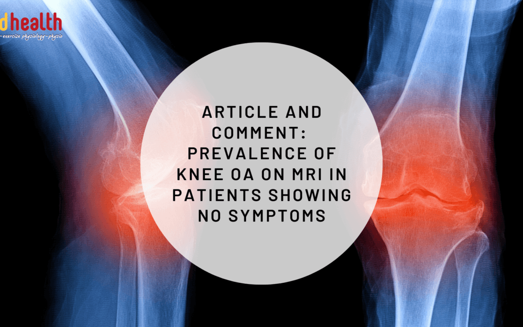
MRI is the most reliable form of medical imaging available. However, MRI and other medical imaging is very costly, running up costs of over $14 billion in the USA annually.
MRI’s often show findings such as osteoarthritic/cartilage changes, meniscal tears, bone marrow lesions and osteophytes, which are often interpreted as causes of pain and symptoms.
These findings often trigger further medical/surgical opinions and interventions, which again drives the costs up for each individual patient. However, more and more recently published studies are deciphering that the relationship between MRI findings and the signs and symptoms of osteoarthritic knee pain is imprecise and often not a true diagnostic indicator.
Culvenor et.al. (2019) recently published a systematic review of the evidence around MRI findings in knees of asymptomatic individuals. The review looked at 5397 knees in total, of 4751 individuals, with the main findings as follows:
- Of these knees, 24% showed cartilage defects
- 10% showed meniscal tears
- In individuals over 40 years of age, these numbers increased to 43% and 19% respectively
- The overall prevalence of osteoarthritic changes in these knees was 4-14% in those under 40 years old, rising to 19-43% in those over 40.
What do these results tell us?
Since more than one-third of the older population will exhibit osteoarthritic features in knee MRI’s, medical and/or surgical interventions targeting these imaging findings may NOT alleviate pain in patients with knee symptoms.
What should I do if I have a knee injury?
The best thing to do for any knee injury is consult a Physiotherapist or Exercise Physiologist initially, and get a diagnosis based on your symptoms and clinical signs. If symptoms are severe enough, these professionals may refer for imaging, but will (at least) also try some exercise-based rehabilitation for the injury and recommend sticking with this routine for at least 3 months. If you haven’t had a scan, have been doing specific resistance-based exercises for at least 3 months, AND if pain persists, medical imaging may be indicated to lead to further specialist/surgeon intervention. This is the most cost-effective and evidence-based approach to injury rehabilitation.
Want to know more?
If you want more information regarding this article or would like to book for a FREE full body assessment with one of our Physiotherapists or Exercise Physiologists, call us on 9857 0644 or email us at admin@md-health.com.au
Prevalence of knee osteoarthritis features on magnetic resonance imaging in asymptomatic uninjured adults: A systematic review and meta-analysis
Knee MRI is increasingly used to inform clinical management. Features associated with osteoarthritis are often present in asymptomatic uninjured knees; however, the estimated prevalence varies substantially between studies. We performed a systematic review with meta-analysis to provide summary estimates of the prevalence of MRI features of osteoarthritis in asymptomatic uninjured knees.


